In The News

16 May 2024
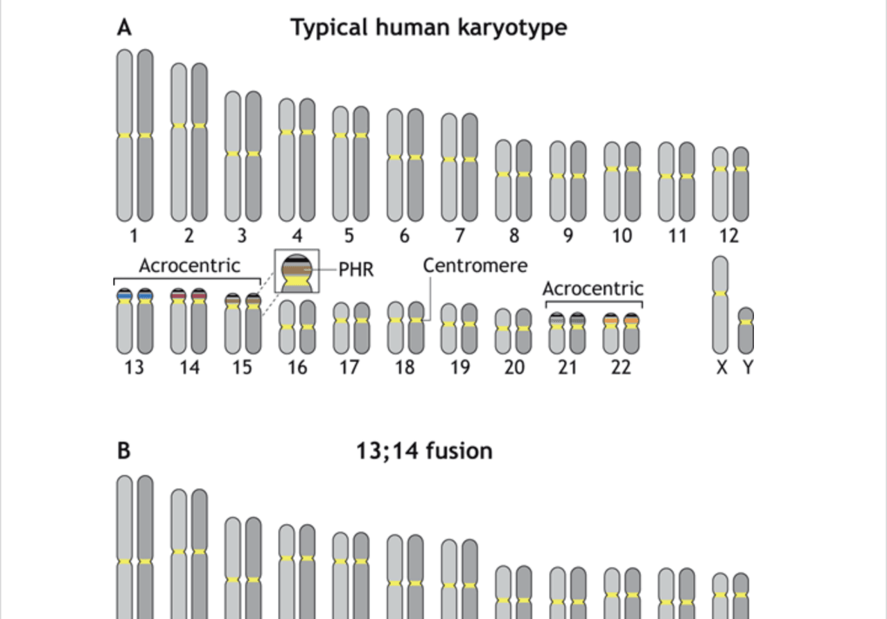
A working model for the formation of Robertsonian chromosomes
From The Journal of Cell Science, read a hypothesis from Stowers Investigator Jennifer Gerton, Ph.D., about the formation of Robertsonian chromosomes.
Read Article
News
Stowers scientists develop visual tool providing insight into the regulation of protein-producing ribosomes and how faulty ribosomes may lead to human disease

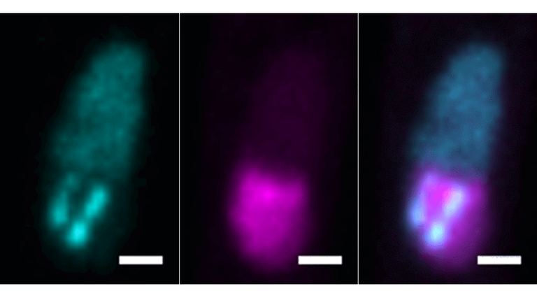
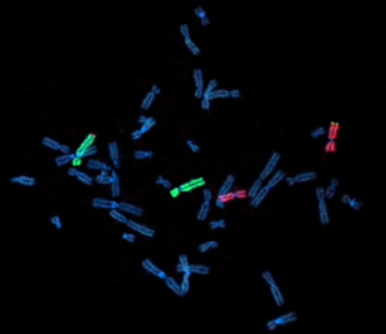
GapR-GFP is a new visual tool for analyzing the spatial organization of ribosomal DNA.
Essential to nearly every process in the body, proteins are encoded by our genes and made in every cell by molecular machines called ribosomes. Generating ribosomes is an energy-consuming process that needs to be carefully regulated to ensure cells have the appropriate amount based on factors such as growth state, nutrient availability, and environmental signals.
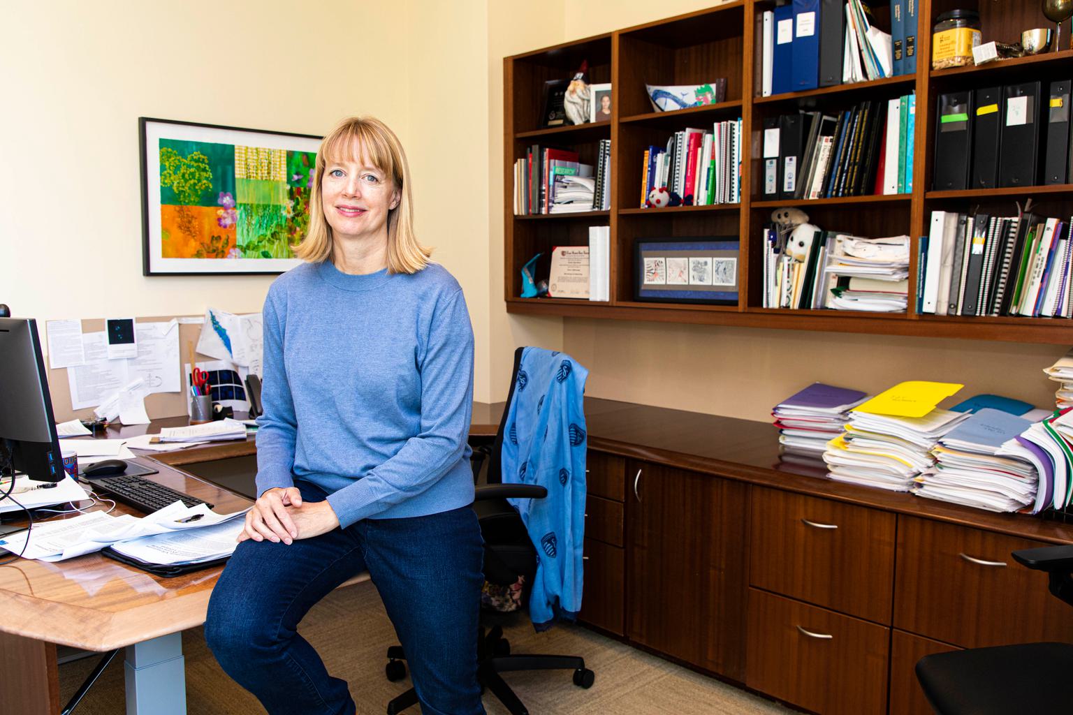
A research article from the Stowers Institute for Medical Research published in PLoS Genetics on July 5, 2024, describes the development of a new visualization tool to examine the spatial organization of ribosomal DNA, regions of DNA involved in the generation of ribosomes. The research team, led by Alexandria Cockrell, Ph.D., from the laboratory of Investigator Jennifer Gerton, Ph.D., validated the ability of this tool, a fluorescent imaging marker, to correlate specific patterns of ribosomal DNA with changes in ribosome generation and then used it to identify new regulators of this process. The findings could lead to a better understanding of conditions and diseases associated with ribosome abnormalities, such as bone marrow failure and the connected predisposition for blood cancer later in life.
“Our ribosomal DNA marker provides a new tool to study the organization of an essential region of the genome in live cells,” said Cockrell, a member of the M.D.-Ph.D. physician-scientist training program at the University of Kansas Medical Center who performed her Ph.D. thesis research in the Gerton Lab.
“We successfully applied this tool in a genome-wide imaging screen in fission yeast, an ideal research system for studying fundamental cellular processes,” said Cockrell . “We were able to identify gene mutations with previously unknown effects on ribosomal DNA spatial organization.”

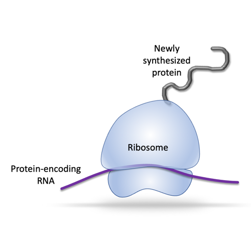
Ribosomes read genetic instructions encoded in RNA messages to produce proteins needed by the cell. Ribosomes themselves are complexes made up of both proteins and RNAs.
Ribosomes produce proteins, yet they are also made up of proteins. Each ribosome is a large complex consisting of approximately 80 different proteins and several RNA molecules encoded by ribosomal DNA. The imaging tool, named GapR-GFP, consists of green fluorescent protein (GFP) fused to a protein that binds to DNA where genes are highly active, particularly those within ribosomal DNA during ribosome production.
Previous studies have examined how changes in ribosome generation correspond to the appearance and structure of the nucleolus, the specialized compartment of the cell nucleus where ribosomes are assembled. Some changes, such as enlargement of the nucleolus, have been identified as predictive indicators for human cancers, which require increased protein production and thus more ribosomes to grow and divide.
Portions of chromosomes containing ribosomal DNA are responsible for creating the nucleolar compartment. These regions contain multiple copies of the genes encoding the RNA components of ribosomes to help meet the cell’s demand. The researchers wanted to determine how the spatial organization of ribosomal DNA is related to its function and to nucleolar structure, a question previously limited by the lack of methods to visualize ribosomal DNA organization.
After introducing GapR-GFP into fission yeast, the researchers conducted proof-of-principle experiments by activating or inhibiting ribosome generation. They found that activation led to visual expansion of both ribosomal DNA and the nucleolar compartment, whereas inhibition resulted in ribosomal DNA condensing and retracting from the nucleolus and compaction of the nucleolus.

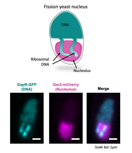
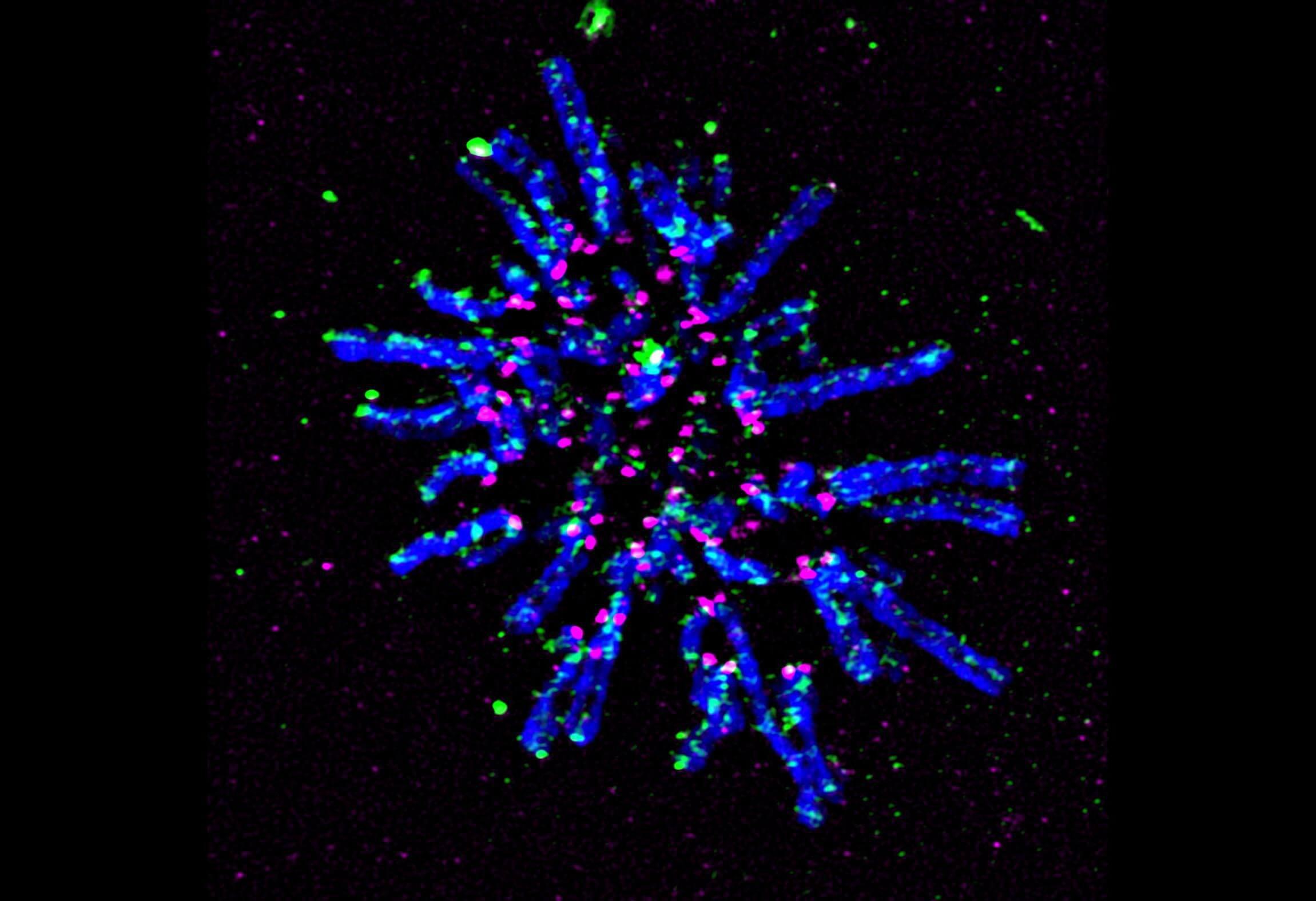
GapR-GFP is a visual tool for analyzing ribosomal DNA in fission yeast. The merged image shows ribosomal DNA (teal) in the nucleolus (magenta).
“Using GapR-GFP, we saw that ribosomal DNA arrays show visual changes based on how many ribosomes the cell needs to make,” said Cockrell.
Next, the researchers introduced GapR-GFP into hundreds of yeast strains having unique genetic mutations. A group of the mutants that showed increased ribosomal DNA size compared to control cells were missing genes for building the ribosome complex.
Further genetic experiments showed that in these ribosomal protein mutants, different genes are activated, indicating a response by the cells to compensate for the reduction of ribosome proteins. Identifying the genes involved in the compensation response gives researchers new modulators to explore in the context of ribosomal DNA regulation and ribosome production.

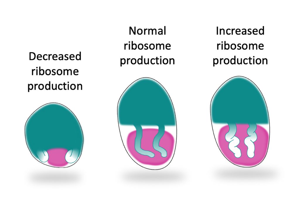
The spatial organization of ribosomal DNA can reflect ribosome production activity. Decreased activity leads to condensation whereas increased ribosome production results in expansion.
“Our work presents the first broad investigation for regulators of ribosomal DNA spatial organization in any organism,” said Cockrell. “Importantly, we have shown that when cells have difficulty producing ribosomes, they may attempt to compensate by using alternative tactics.”
“Our findings have potential implications for human health,” said Gerton. “Ribosomal protein insufficiency occurs in several human disease contexts. A compensation response could contribute to disease processes in cancer and developmental disorders known as ribosomopathies where ribosomal protein insufficiency occurs.”
Additional authors include Jeffrey J. Lange, Ph.D., Christopher Wood, Ph.D., Mark Mattingly, Scott M. McCroskey, William D. Bradford, Juliana Conkright-Fincham, Ph.D., and Lauren Weems from the Stowers Institute, and Monica S. Guo, Ph.D., from the University of Washington School of Medicine, Seattle.
This research was funded by a training grant from National Institutes of Health (NIH) (award: F30GM140716 to AJC), a grant from the National Institute of General Medical Sciences of the NIH (award: R00GM134153 to MSG), and institutional support from the Stowers Institute for Medical Research. The content is solely the responsibility of the authors and does not necessarily represent the official views of the NIH.
In The News
16 May 2024
From The Journal of Cell Science, read a hypothesis from Stowers Investigator Jennifer Gerton, Ph.D., about the formation of Robertsonian chromosomes.
Read Article
News

03 April 2024
Collaborative study helps enlighten a paradox regarding chromosome transmission, potentially leading to a greater understanding of how variations in these regions underlie genome instability and disease
Read Article
News

29 February 2024
"We hope to provide insights into the basic molecular functions of these genes that can someday be harnessed to help people with mutations and their families."
Read Article
News

04 December 2023
Centromeres—specific regions of DNA typically located near the center of chromosomes—achieve a common core structure to ensure proper alignment and segregation of chromosomes during cell division.
Read Article
