News
15 April 2025
Light sheet microscopy: A decade-long journey from DIY innovation to cutting-edge imaging
A look at the technology that provides researchers with deeper insights into complex biological systems.
Read Article
Stowers scientists show how pluripotent stem cells mobilize in wounded planarian worms, to better understand stem cell behavior in regeneration and disease
KANSAS CITY, MO—The skin, the blood, and the lining of the gut—adult stem cells replenish them daily. But stem cells really show off their healing powers in planarians, humble flatworms fabled for their ability to rebuild any missing body part. Just how adult stem cells build the right tissues at the right times and places has remained largely unanswered.

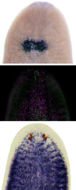
After transplantation of healthy tissue and subsequent decapitation, planarian stem cells (shown in blue, top panel) leave the graft (shown in green, middle panel) and migrate towards the amputation site while proliferating and producing progeny (magenta). This wound-induced migration rescues the lethally irradiated host animal and eventually the stem cell compartment is completely repopulated with fully functional stem cells (shown in blue, bottom panel).
Image: Courtesy of Dr. Otto C. Guedelhoefer, IV, University of California, Santa Barbara
Now, in a study published in upcoming issue of Development, researchers at the Stowers Institute for Medical Research describe a novel system that allowed them to track stem cells in the flatworm Schmidtea mediterranea. The team found that the worms’ stem cells, known as neoblasts, march out, multiply, and start rebuilding tissues lost to amputation.
“We were able to demonstrate that fully potent stem cells can mobilize when tissues undergo structural damage,” says Howard Hughes Medical Institute and Stowers Investigator Alejandro Sánchez Alvarado, Ph.D., who led the study. “And these processes are probably happening to both you and me as we speak, but are very difficult to visualize in organisms like us.”
Stem cells hold the potential to provide an unlimited source of specialized cells for regenerative therapy of a wide variety of diseases but delivering human stem cell therapies to the right location in the body remains a major challenge. The ability to follow individual neoblasts opens to door to uncovering the molecular cues that help planarian stem cells navigate to the site of injury and ultimately may allow scientists to provide therapeutic stem cells with guideposts to their correct destination.
“Human counterparts exist for most of the genes that we have found to regulate the activities of planarian stem cells,” says Sánchez Alvarado. “But human beings have these confounding levels of complexity. Planarians are much simpler making them ideal model systems to study regeneration.”
Scientists had first hypothesized in the late 1800s that planarian stem cells, which normally gather near the worms’ midlines, can travel toward wounds. The past century produced evidence both for and against the idea. Sánchez Alvarado, armed with modern tools, decided to revisit the question.
For the new study, first author Otto C. Guedelhoefer, IV, Ph.D., a former graduate student in Sánchez Alvarado’s lab, exposed S. mediterranea to radiation, which killed the worms’ neoblasts while leaving other types of cells unharmed. The irradiated worms would wither and die within weeks unless Guedelhoefer transplanted some stem cells from another worm. The graft’s stem cells sensed the presence of a wound—the transplant site—migrated out of the graft, reproduced and rescued their host. Unlike adult stem cells in humans and other mammals, planarian stem cells remain pluripotent in fully mature animals and remain so even as they migrate.
But when Guedelhoefer irradiated only a part of the worm’s body, the surviving stem cells could not sense the injury and did not mobilize to fix the damage, which showed that the stem cells normally stay in place. Only when a fair amount of irradiated tissue died did the stem cells migrate to the injured site and start to rebuild. Next, Guedelhoefer irradiated a worm’s body part and cut it with a blade. The surviving stem cells arrived at the scene within days.
To perform the experiments, Guedelhoefer adapted worm surgery and x-ray methods created sixty to ninety years ago. “Going back to the old literature was essential and saved me tons of time,” says Guedelhoefer, currently a postdoctoral fellow at the University of California, Santa Barbara. He was able to reproduce and quantify results obtained in 1949 by F. Dubois, a French scientist, who first developed the techniques for partially irradiating planarians with x-rays.
But Guedelhoefer went further. He pinpointed the locations of stem cells and studied how far they dispersed using RNA whole-mount in situ hybridization (WISH), specifically adapted to planarians in Sánchez Alvarado’s lab. Using WISH, he observed both original stem cells and their progeny by tagging specific pieces of mRNA . The technique allowed him to determine that pluripotent stem cells can travel and produce different types of progeny at the same time.
“In other systems, most migrating stem cell progeny are not pluripotent,” says Guedelhoefer. “For the most part, blood stem cells in humans stay in the bone marrow but their progeny leave and turn into a few other cell types.” But in planarians, it looks like those two things are completely separate. Stem cells can move and maintain the full potential to turn into other types of cells.”
Next, Sánchez Alvarado looks forward to implementing genetic screens and transplantation experiments to disrupt or enhance the cellular behaviors the team observed, to figure out the “rules of engagement” for stem cell migration, he says.
“Why can some animals regenerate whole body parts but you and I are not good at it?” says Sánchez Alvarado. “Can we write an extra rule or erase one? Is it possible, for instance, to get rid of cancer while gaining regenerative properties? These are questions we’d love to have answers to.”
The Stowers Institute for Medical Research, a National Institutes of Health Training Grant (5T32 HD0791), a National Institutes of Health Grant (GM057260), and Howard Hughes Medical Institute provided funding for this work
News
15 April 2025
A look at the technology that provides researchers with deeper insights into complex biological systems.
Read Article
News
11 April 2025
“There are few rewards as powerful and as elevating as making a clear, robust scientific observation that advances the field.”
Read Article
News

09 April 2025
New study shows how we can better learn our genome’s hidden grammar, potentially paving the way for personalized medicine.
Read Article
