News
15 April 2025
Light sheet microscopy: A decade-long journey from DIY innovation to cutting-edge imaging
A look at the technology that provides researchers with deeper insights into complex biological systems.
Read Article
Press Release
New research from the Stowers Institute for Medical Research has identified a backup for an important biological system – the hematopoietic system, whose adult stem cells constantly replenish the body’s blood supply.

KANSAS CITY, MO—New research from the Stowers Institute for Medical Research has identified a backup for an important biological system – the hematopoietic system, whose adult stem cells constantly replenish the body’s blood supply.
The finding provides evidence that hematopoietic stem cells (HSCs) consist of different functional populations of cells, including “primed” cells, which are ready at a moment’s notice to be activated to produce red blood cells, white blood cells, and platelets, and “reserve” cells, which come into play when there has been damage to the first-line stem cells.
“Under severe stress – radiation or chemotherapy that injures blood cells, including primed hematopoietic stem cells – that’s when reserve cells kick in,” says Linheng Li, Ph.D., an Investigator at the Stowers Institute and senior author of the study.
Reserve HSCs could hold the key to understanding how blood cells repopulate after severe injury depletes the bone marrow that produces blood cells. This concept may provide an insight for understanding treatment-resistant leukemia cells. The study appears online January 15, 2019, in the journal Cell Reports.

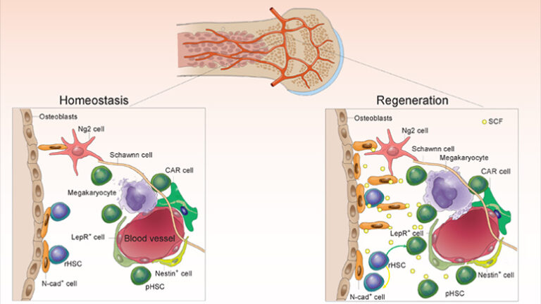
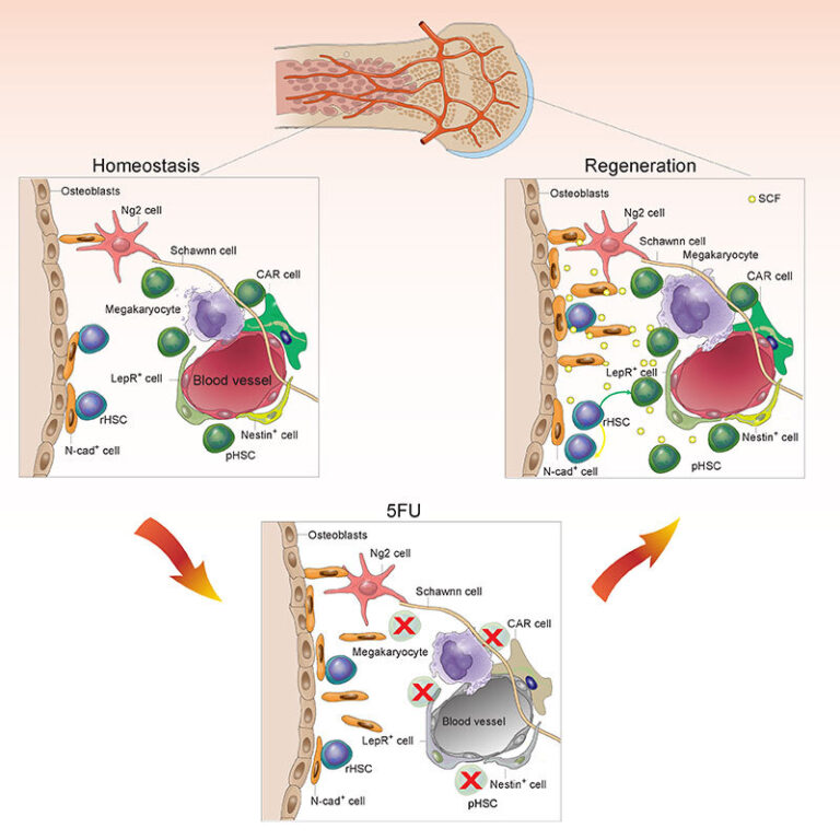
Co-existence of rHSCs and pHSCs in bone marrow with the latter sensitive to chemotherapy but the former surviving chemotherapy and restoring the HSC pool
Image: Courtesy of Mark Miller
Every second, two million new blood cells are churned out by the amazing, regenerative HSCs that reside in the core of most bones. Only a fraction of these cells actively move through the cell cycle at any given time; the rest lie in an inactive or quiescent state, which was once believed to protect them from harm. However, several studies have shown that the majority of these quiescent HSCs are sensitive to DNA damage from chemotherapy.
Over a decade ago, Li suggested that a special population of HSCs that are resistant to damage might still reside in the bone marrow, hidden in some unexplored niche. In this study, Meng Zhao, Ph.D. (now a faculty member at Sun Yat-Sen University in Guangzhou, China), Fang Tao, Ph.D., Zhenrui Li, Ph.D. (now a fellow at St. Jude Children’s Research Hospital), Aparna Venkatraman, Ph.D., and Xi C. He, M.D., undertook a series of experiments using a mouse model to prove that these cells exist.
A key experiment utilized a cell surface marker to isolate reserve and primed HSCs. The researchers transplanted reserve HSCs or primed HSCs into recipient animals. After the engraftments were established, they treated recipient mice with the chemotherapy agent 5-fluorouracil (5-FU). While the primed HSCs reflected by their derived blood cells began to decline after treatment, the reserve HSCs’ derivatives were unscathed. “In effect, we showed that hematopoietic stem cells have functionally distinct subpopulations – one that acts under normal conditions, and the other that acts under times of stress,” says Li.
Next, the researchers examined bone and marrow from transplanted mice, labeling the reserve cells with fluorescent tags and studying them microscopically to pinpoint their location in the bone marrow. With Sarah Smith, Ph.D., from the Stowers Microscopy Center, they spotted the fluorescently labeled cells lurking in a specialized niche along the inside surface of the bone, adjacent to a population of cells known as N-cadherin+ bone-lining cells, which was reported by the Li team in 2003 as the first niche identified to support HSCs. In the current study, Tao and colleagues found that these N-cadherin+ bone-lining cells are mesenchymal or skeletal stem cells with the potential to produce bone, cartilage, and fat. Therefore, N-cadherin may be used as a marker to isolate skeletal stem cells and may have potential uses in bone and cartilage regenerative medicine.
These N-cadherin+ bone-lining cells appear to protect the reserve cells from injury by feeding them survival factors like the aptly named stem cell factor and others. When the researchers depleted these support cells, the reserve cells were no longer able to survive chemotherapy treatment.
“Interestingly, we found that N-cadherin+ cells lining the bone are also resistant to chemotherapy, while stromal cells in the central marrow are sensitive to chemotherapy, which allows N-cadherin+ cells to better support the reserve stem cells,” said Tao, who, along with Zhao, is co-first author on the study.
Other coauthors include Jay Unruh, Ph.D., Shiyuan Chen, Christina Ward, Pengxu Qian, Ph.D., John M. Perry, Ph.D., Heather Marshall, Ph.D., and Jinxi Wang, M.D., Ph.D.
The work was supported by funding from the Stowers Institute for Medical Research to L.L. and a grant to the University of Kansas Cancer Center from the National Cancer Institute of the National Institutes of Health under award number P30CA168524. Additional support included National Key Research and Development Program of China (2017YFA0103403, 2018YFA0107203) to M.Z.; Department of Biotechnology, Ministry of Science and Technology, Government of India overseas associateship to A.V.; and the National Institutes of Health award numbers R01AR059088 and R01DE018713 to J.W. The content is solely the responsibility of the authors and does not necessarily represent the official views of the National Institutes of Health.
Lay Summary of Findings
Blood-forming adult stem cells reside deep in the bone marrow and are responsible for regenerating the body’s blood supply including red blood cells, white blood cells, and platelets. In a study published online January 15, 2019, in the journal Linheng Li, Ph.D., and collaborators investigated how these cells, despite being sensitive to DNA damage, manage to repopulate blood cells after chemotherapy or injury has depleted their numbers.
The researchers report that a subset of blood-forming, or hematopoietic, adult stem cells called “reserve” hematopoietic stem cells (HSCs) are resistant to chemotherapy. These reserve HSCs are located in a specialized niche, composed of cells on the inner bone surface expressing the molecule N-cadherin, in mouse bone marrow. The finding advances the understanding of HSC biology and may open new avenues for treating blood diseases like leukemia and autoimmune disorders.
About the Stowers Institute for Medical Research
Founded in 1994 through the generosity of Jim Stowers, founder of American Century Investments, and his wife, Virginia, the Stowers Institute for Medical Research is a non-profit, biomedical research organization with a focus on foundational research. Its mission is to expand our understanding of the secrets of life and improve life’s quality through innovative approaches to the causes, treatment, and prevention of diseases.
The Institute consists of 17 independent research programs. Of the approximately 500 members, over 370 are scientific staff that include principal investigators, technology center directors, postdoctoral scientists, graduate students, and technical support staff. Learn more about the Institute at www.stowers.org and about its graduate program at www.stowers.org/gradschool.
Media Contact:
Joe Chiodo, Head of Media Relations
742.462.8529
press@stowers.org
News
15 April 2025
A look at the technology that provides researchers with deeper insights into complex biological systems.
Read Article
News
11 April 2025
“There are few rewards as powerful and as elevating as making a clear, robust scientific observation that advances the field.”
Read Article
News

09 April 2025
New study shows how we can better learn our genome’s hidden grammar, potentially paving the way for personalized medicine.
Read Article
