By Maria Vacek Broadfoot
Stowers Institute amasses menagerie of unconventional organisms to study health and disease.
When it comes to the intricacies of biology, humans are a challenging species to study. Thankfully, scientists can seek insight elsewhere in the animal kingdom. Our planet is home to an estimated 30 million species of animals, ranging from rodents that weigh less than a teaspoon of sugar to whales with hearts the size of VW Beetles. Although we look very different on the outside, our biology is often more similar than different, especially at the level of cells and genes.
Over many decades, biologists have brought several of these organisms into their laboratories, assembling aquariums for striped zebrafish or vials to house skittering fruit flies. However, they have mostly stuck with a small handful of animal models, favored because they are easy to domesticate or are simple to breed in large numbers.
Robb Krumlauf, PhD, thinks it is time to branch out. As scientific director of the Stowers Institute, he is not surprised that Stowers investigators are amassing a menagerie of emerging model organisms, including bloodsucking lamprey eels, blind cavefish, self-regenerating flatworms, and fluorescent sea anemones.
"The ability to mine the diversity of nature in a mechanistic way is a real strength of modern biology," says Krumlauf. "Our philosophy is to use the latest molecular tools to ask specific questions about the unique way other organisms do things, and to discover new biology that will hopefully be important for understanding human disease," says Krumlauf.
Bloodsucking lamprey eels
Krumlauf knows a bit about developing model organisms. As a young scientist in the 1980's, he was among the first to insert genes into the mouse genome to create "transgenic" mice that mimic human development and disease. Since then, he has used these mouse models to study how vertebrates like mice and humans evolved their unique features. His work in mice has shown that a set of genes, called Hox genes, control the layout of a developing embryo, marking where structures should appear along the bodyplan from head to tail. This sort of molecular ruler is present not only in mice and humans, but also at play in more primitive organisms, from flies, to worms, to fish.
Further down the tree of life, back 500 million years to the base of the vertebrate branch, lies one of the most unappealing animals in existence, the sea lamprey Petromyzon marinus. These long, eel-like fish use a jawless mouth armed with spiraling rows of horny teeth to latch on to their prey and feed. Researchers had assumed lamprey lacked jaws because their Hox genes weren't operating the same way as they were in higher vertebrates. Krumlauf thought this jawlessness might be due to the activity of Hox genes in a specialized brain structure called the hindbrain, which coordinates basic functions like heartbeat, breathing, and jaw movement. But testing this theory meant Krumlauf would have to get his hands on these bloodsucking creatures.
Though lamprey are difficult to study in captivity, there are plenty of these parasitic fish in the sea. Just like adult salmon return to the spawning ground of their birth, lamprey swim from the ocean to freshwater streams and lakes to spawn and lay their eggs. Every summer, Krumlauf's collaborator, Marianne Bronner, PhD, obtains egg-bearing lampreys out of the waters of the Great Lakes and sets them up in tanks at CalTech, where the slimy sea creatures churn out tens of thousands of fertilized eggs. At the same time, Stowers researchers migrate from Kansas City to California each summer to study the resulting embryos—tiny translucent spheres that belie their horny-toothed parentage.
"I don't think any of us would look forward to studying this animal if we had to work with the full grown organisms all the time. Lamprey are the ugliest beasts you can possibly imagine. But the embryos are really striking and beautiful," says Krumlauf.
During one of these trips, a member of Krumlauf's team, Hugo Parker, PhD, manipulated these tiny embryos by attaching fluorescent tags to the molecular switches that turn the Hox genes on or off. They then placed each embryo under a fluorescence microscope and watched the light show unfold as the organism began to develop towards its adult form. To their surprise, the molecular switches in the hindbrain lit up exactly the same way in jawless lamprey as they did in jawed vertebrates like zebrafish. So how could the same genes give rise to such strikingly different animals?
"We think it may be like going to the hardware store, where you can buy the same building materials but you can build a lot of different kinds of structures with the same tools and materials," says Krumlauf. "The ancestral function of the original genes might be the same, but these organisms have evolved different functions over the years."
Now, the Krumlauf team is studying the mechanisms driving this evolutionary process, which they hope will give insight into how these instructions are used in humans and how they can get misinterpreted in disease.
Blind cavefish
Unlike the lamprey, which has a face only a mother (or dedicated scientist) could love, the cavefish Astyanax mexicanus is a popular aquarium oddity. These creatures not only lack eyes but are also completely devoid of pigment, giving their bodies an almost ghostly pinkish-white sheen. Cavefish are more than just a funny-looking pet – they are the product of evolutionary forces at their most brutal and unforgiving.
In the wild, these cave dwellers must survive in pitch-black surroundings, where the only food available is swept into the caves once a year when rivers flood. Along with animal and plant remains, the floods also bring in fresh recruits of surface fish from nearby waters that must also adapt to their harsh new environment, or die trying. Over time, the gene pool of the survivors evolves as they shed seemingly pointless properties, like eyesight and coloring, and acquire vitally important ones, like starvation resistance.
Assistant Investigator Nicolas Rohner, PhD, studies this case of extreme evolution, and has found cavefish remain startlingly healthy despite enduring repeated cycles of feast or famine. When floods finally deliver their annual meal, the ravenous cavefish gorge themselves until all the food is gone. By the time they are done eating, many of their organs have doubled in size and they have become ten times fatter than their surface counterparts. Yet these fleshy fish are not prone to any of the obesityrelated conditions that plague humans.
"They are a great model for understanding why we as a species store so much fat," says Rohner, who recently completed a postdoc at Harvard and launched his own laboratory at the Institute. "The typical human has about 25 percent body fat; our closest living ape relatives have much less than one percent. From an evolutionary perspective, humans carry a significant amount of body fat, and with modern diets, it's only increasing. It is important to understand why humans are prone to obesity or why we develop illnesses like diabetes and heart disease."
As a postdoc, Rohner maintained tanks full of fat-filled cavefish and svelter, albeit more mundane-looking, surface fish. By breeding the two types of fish with each other, he could follow the inheritance of various traits across generations, much like Gregor Mendel did with his famed pea plants. Rohner combined this classical genetic approach with modern sequencing technology to pinpoint regions of the genome associated with feeding behaviors and appetite. He found cavefish have a mutation in melanocortin 4 receptor, or MC4R, a gene known to give people an insatiable appetite when mutated. It is the most common single genetic cause of obesity.
Now that he is at the Stowers Institute, Rohner and his team will be looking for mutations to explain why cavefish are able to store so much fat without suffering the negative consequences. Such knowledge could lead to new treatments to buffer the effects of metabolic diseases.
"The link between evolution and medicine is undeniable," says Rohner. "Diseases have an evolutionary underpinning, so understanding why we are the way we are could help us to find new approaches to address our weaknesses."
Self-regenerating flatworms
For researchers like Rohner and Krumlauf, tapping the animal repertoire outside of typical lab models allows them to study specific characteristics in their most extreme, exaggerated forms. Gluttonous cavefish become the perfect animal for examining human metabolism. Jawless lamprey present an excellent model for investigating the complexities of brain and head development. Likewise, the self-regenerating flatworm embodies the ideal system for understanding how some organisms regrow damaged organs or missing body parts.
"The technological powers in biology are unprecedented – in a matter of days you can literally go from knowing nothing about an organism to having a comprehensive list of all of the genes that are turned on or off," says Investigator Alejandro Sánchez Alvarado, PhD. "We are witnessing the future of biomedical research, where advances like sequencing and genome editing mean we are no longer constrained to domesticated animals but can look elsewhere in nature to answer questions about disease, aging, and regeneration."
Sánchez Alvarado believes regeneration is one of the last untamed frontiers of developmental biology. Despite decades of research, scientists still don't understand how a few lucky members of the animal kingdom manage to perform this amazing feat. Salamanders can grow back a pinched tail; zebrafish can regrow fins or even chunks of damaged heart tissue. One of the most talented of these regenerative magicians, planaria, can grow an entirely new head after being decapitated. Cutting these miniscule, arrow-shaped flatworms into pieces results in a growing brood of full-sized clones.
Twenty years ago, Sánchez Alvarado began reading up on these resilient creatures, eager to learn their secrets. When he learned that the planaria Schmidtea mediterranea, which possesses a small genome size and a small number of chromosomes, could be obtained from an abandoned fountain in a park in Barcelona, he and his then post-doctoral fellow Phil Newmark, PhD, flew to the city and set liver-baited traps in every fountain they could find. The researchers then turned their catches into a line of hundreds of thousands of planaria that are now being used in laboratories all across the United States.
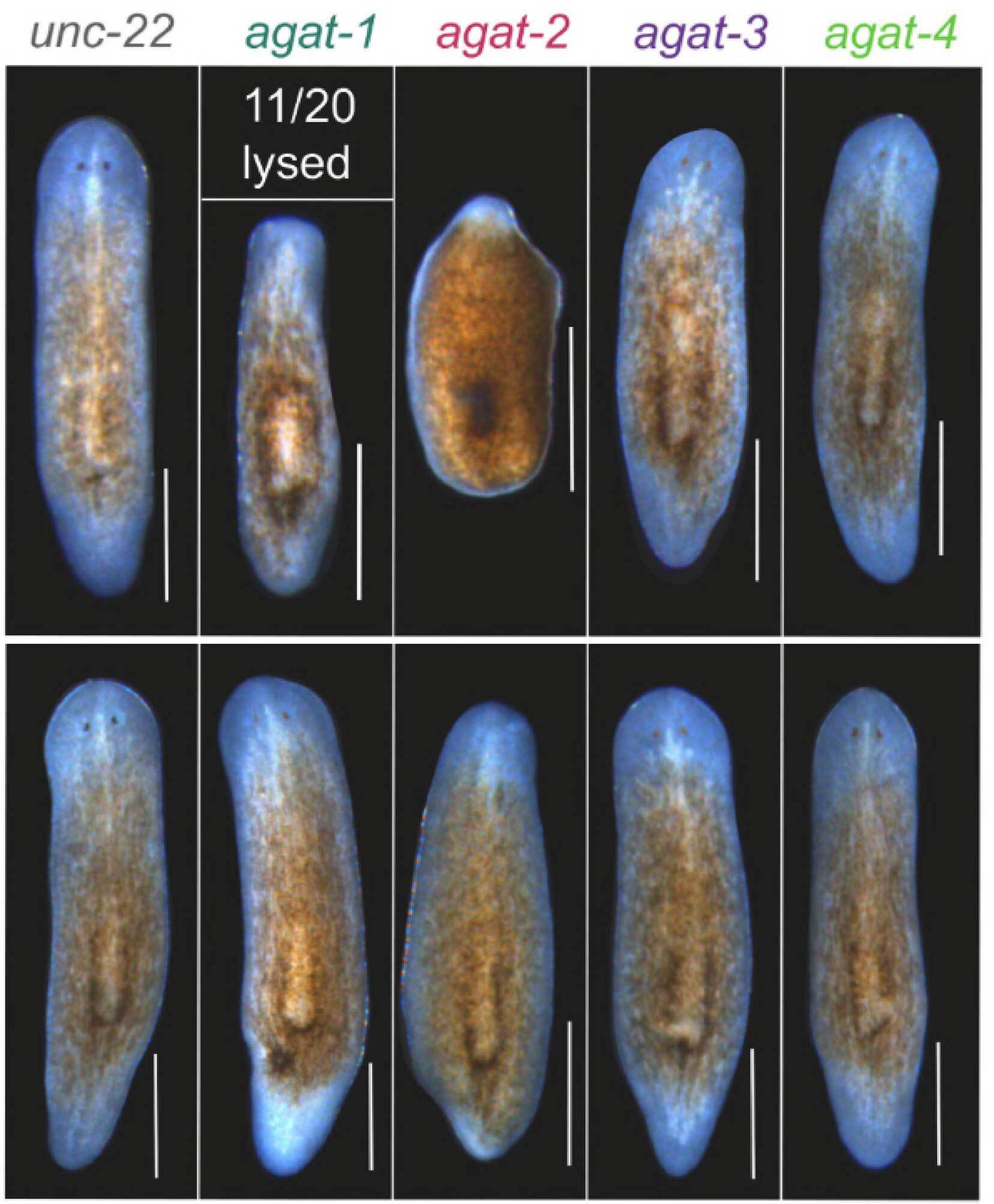
In Sánchez Alvarado's own laboratory, his team is creating a genetic flow chart to describe how this miraculous process unfolds in planaria. First, they take a razor blade to the animal, cutting off a head or vital organ like the pharynx. Then they measure how the worm's more than 20,000 genes are turned on or off as they begin to grow back pieces of flesh. Once the researchers know which genes are at play, they use advanced molecular techniques like RNA interference to silence each of these genes to see how it affects the animal's ability to regenerate the next time it goes under the knife.
Thus far, his team of scientists has identified several groups of genes responsible for successful regeneration. Some, like a gene called ß-catenin, help the organism figure out whether it needs to grow back a head or a tail. Others, including the gene FoxA, are in charge of rebuilding a specific organ. Ultimately, each disfigured planaria is made whole again through the action of stem cells, the same cellular entities responsible for replenishing dead or damaged cells that course through our veins, line our gut, and cover our skin.
"If we could glean information about conserved mechanisms between worms and humans, we could understand why we can only use these same cells to repair simple wear-and-tear and not to launch a full-blown regenerative response," says Sánchez Alvarado.
Unfortunately, nobody knows whether regeneration is an ability other organisms evolved separately or if it is something that the human lineage once had and lost. To answer that question, researchers will have to examine closely our evolutionary ancestors to find clues for potential human self-healing.
Fluorescent sea anemones
Analyzing the genetic underpinnings of a wide variety of animal species is getting easier as sequencing becomes cheaper and more routine. At last count, the genomes of nearly 2,500 multicellular organisms had been sequenced, including sea lampreys, cavefish, planaria, and sea anemones. These projects have given some unexpected surprises. Take the starlet sea anemone, Nematostella vectensis, a seemingly primitive animal that shares a phylum with other simpletons like corals and jellyfish. Unexpectedly, its genome was found to be large and complex, sharing more in common with humans and other vertebrates than traditional model organisms like fruit flies or roundworms.
This incongruous finding attracted the attention of Associate Investigator Matthew Gibson, PhD, who has spent most of his career studying development in the fruit fly. Gibson is interested in how animal cells become stacked into highly organized layers called epithelia. These epithelial sheets line almost all body surfaces, constructing barriers for body walls, tissues, and organs. Nematostella turned out to be a stunning example of this type of cellular organization, an organism Gibson describes as a "beautiful bag of epithelium."
Close up the sea anemone is strikingly beautiful, a translucent, fluid-filled stem that is crowned with over a dozen delicate tentacles like the petals on a flower. But the organism is hardier than it looks. It doesn't seem to die of natural causes, and will even survive being cut in two by regenerating its other half. When Gibson and his team first viewed these sea anemones under a fluorescence microscope, they noticed glowing patches of bright red color just below the animal's tentacles. Out of curiosity, they cloned the party responsible for the mysterious red fluorescence, a gene later named NvFP-7R.
Researchers don't know exactly what purpose NvFP-7R serves in sea anemone, though some think it might act as a sort of sunscreen that protects the organism from harmful rays. Gibson and colleagues came up with their own use for the fluorescent protein, as a target for testing two revolutionary genome-editing tools. They used techniques, known as CRISPRs and TALENs, to "knock-out" the NvFP-7R gene to erase the animal's bright red patches. Now similar experiments are underway to knock out the genes they are most interested in – those involved in controlling development and the arrangement of epithelial cells.
Epithelial cell biology can provide a valuable window into both normal development and the origins of cancer. Epithelial cells make up the vast majority of human cancers — any diagnosis that carries the word "carcinoma" has epithelial roots, and any cancer that has spread did so by escaping the confines of epithelial sheets. While focusing on fundamental cellular, developmental, and evolutionary problems, Gibson emphasizes the potential significance of this work to cancer research.
Like other researchers who share company with unusual organisms such as lamprey, cavefish, and flatworms, Gibson is quick to point out that the results with the biggest impact often come from unexpected places. In fact, the CRISPR system of gene editing has revolutionary biomedical implications, yet was originally discovered by researchers studying how bacteria chop up invading viruses. Today it is being used to cut and paste the genomes of many species at will.
"It is a mistake to think we already know everything there is to know because we have looked at a handful of lab animals in detail," says Gibson. "One of the best ways to learn as much as we can is to look in places no one has looked before or to try to ask questions no one has asked before. Part of gaining access to new knowledge is studying unconventional animal models."


