News
15 April 2025
Light sheet microscopy: A decade-long journey from DIY innovation to cutting-edge imaging
A look at the technology that provides researchers with deeper insights into complex biological systems.
Read Article
News
A look at the technology that provides researchers with deeper insights into complex biological systems.
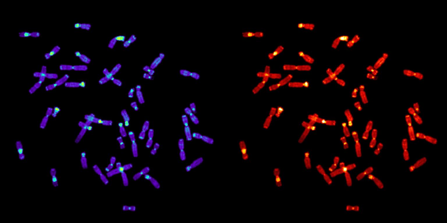
Vibrant pinks and blues that appear to be the structural parts of a fish-like organism are, in fact, genes visible inside individual cells of an entire zebrafish.
"Seeing is believing," explained Zulin Yu, Ph.D., Head of Light Microscopy at the Stowers Institute. "With this system, we can capture fine details of large biological structures in three dimensions.”
The Microscopy Technology Center’s new piece of equipment —a light sheet microscope—is providing researchers and labs with significantly enhanced imaging capabilities.
Achieving incredibly high resolution of a large sample, like a whole organism, is the light sheet fluorescence microscope’s superpower. Conventionally, high resolution and large samples were mutually exclusive.
“So, for example, when you look at a Google map, you can see what a city looks like,” Yu said. “But you can’t look at an individual house or street in fine detail and in three dimensions at the same time. You either lose the house details, or you lose the bigger city picture.”
This isn’t the first time, however, that the microscopy team has used a version of such a scope. Models like the one recently acquired weren’t commercially available in the past, but that did not deter the team.
Around 2013, Sean McKinney, Ph.D., Head of Computational Imaging and Amanda Kroesen, an Associate Scientist in the Microscopy Center, came across research papers describing a new, exciting way to image and saw its potential for advancing research at the Institute. “We thought, this is something we have a skill set for,” she explained.
The pair set out to build their own light sheet scope with published research as their guide. At the time, only a handful of labs worldwide had the expertise and resources to attempt such a project. It worked.
“I probably spent about 10 years doing experiments on that old microscope,” said Kroesen, “And it was a difference-maker on some research projects.”
But advances in technology eventually outpaced what they were able to do with the DIY scope. The home-built system is manually operated, requiring meticulous laser alignment and manual sample loading. It was time for an upgrade.
“I’ll be a little sad to see it taken apart,” said Kroesen.
With the new system, everything is streamlined, automated and user-friendly.
“One of our most important jobs here is training postdocs and students, and sometimes, Investigators,” Yu explained. “Once the user gets an exciting result, they want to use the light sheet system by themselves. We couldn't do that with the old scope, but now researchers can gain expertise and independence.”
In addition to being user-friendly, the new system produces significantly higher quality data and in much less time, it can attain the level of resolution conventionally seen in electron microscopy.
The technology is helping the Özel Lab map the neural connections in the fruit fly brain. And the Kostova Lab can use the scope to see where genes in single cells are distributed throughout a zebrafish embryo.
Further, the laser configuration minimizes photo toxicity so live specimens can be imaged and there’s no need to thin-section a sample. Because the scope allows samples to be imaged in a more natural state, the Sánchez Alvarado Lab can study intercellular communication in flatworms.
"Technology shifts every five to ten years," said Yu. "Our goal is to provide the best tools to our researchers, helping them unlock new scientific discoveries."
News
15 April 2025
A look at the technology that provides researchers with deeper insights into complex biological systems.
Read Article
News
11 April 2025
“There are few rewards as powerful and as elevating as making a clear, robust scientific observation that advances the field.”
Read Article
News

09 April 2025
New study shows how we can better learn our genome’s hidden grammar, potentially paving the way for personalized medicine.
Read Article