22 April 2025
#TechTalk: What is the Cells, Tissues, and Organoids Technology Center?
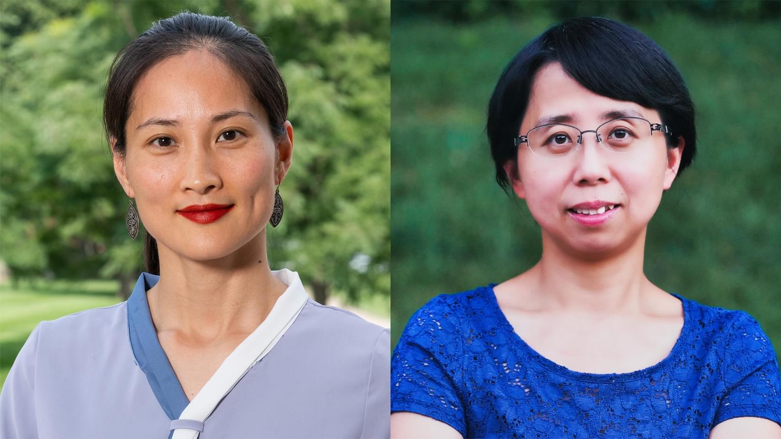
A Q&A with Chongbei Zhao, Director, Cellular, Tissue, and Molecular Biology, and Yan Wang, Scientist I
Read Article
By Nicholas Gerbis
When a protein folds properly, it carries out normal tasks without a hitch. But when it folds into the wrong conformation, as occurs in many prion diseases, the result not only poses a deadly hazard— it also spreads exponentially to impact sister proteins.
While prion proteins can exist in alternative states, they have rarely made headlines except in accounts of their deadly role in brain-destroying disorders like Creutzfeldt-Jakob disease, or their links to the amyloid plaques of Alzheimer’s disease.
Over the past few decades, however, the narrative has shifted to a more nuanced view, rewritten by research revealing that there appear to be normal, functional, even crucial, roles prions can play in cells. At the forefront of these advances stand two teams at the Stowers Institute, one led by Kausik Si, PhD, a pioneer in prion memory research for more than a decade, and the other led by Randal Halfmann, PhD, who brought his new prion model and quantitative methods to the Institute in 2015.
Their challenge lies in developing a clearer picture of how prions choose to adopt one state versus another, why they spread to nearby proteins, and what divides the functional from the dysfunctional. It’s a tall order, but not quite as tall as overcoming the inertia of prevailing scientific wisdom.
A New Frontier
When Randal Halfmann first encountered prions in Susan Lindquist’s lab at Massachusetts Institute of Technology (MIT), he saw a terrain ripe for pioneers.
“It was a wonderful time to get into that field because it was—and it’s really still—a very Wild-West-y kind of a science,” he says.
Halfmann, who grew up on a West Texas cattle ranch, knew a little something about untamed territory. But no run-in with mad cow disease set him on the prion research path. While at Texas A&M, his naturalist bent—which persists today in his hobbies of foraging for wild plants and mushrooms—initially steered him toward plant cytogenetics, the study of plant cells and chromosomes. Prions would wait until graduate school, when a chance meeting with a visiting lecturer lassoed his fancy with the protein folding problem.
Proteins, the building blocks of life, have mastered doing a lot with a little. Though they differ from species to species—even from organ to organ—all proteins draw from the same pool of around 20 amino acids, strung together in electron-sharing links called peptide bonds. This small cast fulfills a seemingly endless list of roles, from transcribing DNA to flexing muscles. This flexibility derives partly from the range of lengths these chains can reach, from a few amino acids to more than 27,000. But the real artistry of proteins, like that of origami, lies in the transformative power of folding.
Yet, even with a complete chemical blueprint and a thorough grasp of the physical laws that govern proteins, scientists often cannot predict how they will fold. Protein ribbons behave less like unwound springs and more like kinked-up garden hoses wrapped in magnets: Some parts repel, others attract and still others swing out of position just as neighboring sections settle into place. Even settling into a nice, low-energy bunch doesn’t guarantee that a protein has reached its true minimum energy fold, or native state.
“This concept of the native state or the native fold can be really misleading,” says Halfmann. “If you start playing around with it and pushing the boundaries, you start finding that there are multiple alternative states with even lower energy wells.”
The protein folding problem, which some of the world’s most powerful computers have tried in vain to solve, instantly captured Halfmann's imagination—and holds it still.
"It’s still a major problem. It was so fascinating, and I just thought, ‘Now there’s a puzzle that is incredibly important.’”
Important is an understatement. Proteins build tissues, catalyze almost all cellular chemical reactions, and move key substances across cell walls. They form the basis of our immune system, hormones, and enzymes. Proteins even control how genes make more proteins.
Properly folded proteins perform these biological assignments successfully. But when proteins fold into the wrong conformation, they can have detrimental effects and may also propagate by forcing identical proteins to fold in their image. These newly minted prions then go and do likewise.
To researchers like Si and Halfmann, such a biological phenomenon seemed too useful for nature to limit it to destructive purposes. It would be like using oxidation-reduction reactions—the world’s principal sources of energy—solely to rust car bumpers.
“Nothing in biology is an accident,” says Halfmann. “Certainly, if it happens, cells find a way to take advantage of it.”
Inspired by this biological adage, Halfmann set out to find more prions and found a lot of them, including examples responsible for aspects of cell signaling, differentiation, and evolution. His yeast studies have proven especially fruitful, turning up two dozen prion-forming proteins that help manage key cellular processes, from transcription (copying DNA data into messenger RNA) to translation (turning messenger RNA sequences into peptides or proteins).
A change in our view of prions was brewing, but new answers also inspired new questions. As Halfmann and his colleagues explored the margin separating functional prions from dysfunctional ones, another problem emerged.
“It was becoming clear that we in the field didn’t have the right conceptual framework for understanding these proteins.”
Fortunately, Halfmann had an idea in mind.
Transitioning to a New Phase
Elementary school science teaches that a phase transition is a shift from one state of matter to another, often involving the release or absorption of energy. It also defines 0°C as the temperature at which water shifts phases from liquid to solid.
In reality, though, the process occasionally requires a little help. Pure water can remain unfrozen even after its temperature drops past its freezing point. Its molecules slow down, wanting to fall into a cozy lattice formation, but can’t quite find the right orientation. Then a tiny energy fluctuation orients some of them into the first microscopic ice crystal that kicks off the process. Once they have it, a wave of freezing sweeps through the liquid almost instantly.
Halfmann says that a similar process might explain prions’ cellular coup.
When cytoplasm, the viscous fluid that holds all of a cell's non-nuclear goodies, becomes supersaturated with prion-forming proteins, it grows unstable, like supercooled water. Drop in a prion, which is in effect a molecular crystal, and it kick-starts a prion formation cascade. The liquid has “frozen.”
As the Halfmann Lab works to prove this hypothesis, it must also address vital limitations in customary prion research methods.
Our grasp of prion proteins has been hindered by a lack of quantitative methods, which rely on statistical analyses of key quantities. Lacking these measurements, early assays relied on color changes in yeast colonies, an indirect method that could only work after 30 or so generations of yeast. Halfmann says that such methods—really extensions of genetic tests used to study diseases—were limited in scope and prone to false positives, and “grossly obscured the real biology.”
“I think that we’re now emerging from that state, or beginning to,” he says. “Now, you can really point at something and say, ‘This is when it’s happening, how much it’s happening,’ and so on.”
Halfmann credits his colleagues for the new evidence, improved technologies, and emerging fields of study that have enabled him to conceive his new model and study it quantitatively.
“The technology that makes the assay possible— robust photoconvertible fluorescent proteins—only recently became available. The timing coincides nicely with a recent frenzy of activity in biophysics and cell biology of liquid-liquid phase separation.”
Thanks for the Memories
The questions these shifts could help answer include ones posed by Halfmann’s Stowers colleague Kausik Si. Si first made a surprising discovery in the prion world in 2003, when he and Eric Kandel, MD, of Columbia University, put forth the idea that a prion protein called CPEB might have a normal role to help the brain form stable long-term memories. In response to a transient electrical signal, they argued, CPEB would switch to a prion state and spread that state to nearby proteins, thereby building neuron links vital to memory.
Today, Si has expanded his research to include cellular memory, in which cells use DNA-binding proteins called transcription factors to “remember” their type and function following cell division. But human memory still occupies a central space in his research.
“How do prions help us form long-lasting memory?” he asks. “If protein aggregates are generally bad for the nervous system, how are some able to perform vital physiological functions?”
Protein aggregates—snowballs built up via “sticky” protein regions—play a vicious role in prion diseases, but this stickiness exists in nonprion proteins, too. According to Shriram Venkatesan, PhD, a postdoctoral researcher in the Halfmann Lab, nearly all cellular proteins contain such regions, but normal folding tucks them away. Venkatesan studies how protein aggregates might help cells by sweeping up misplaced or harmfully plentiful proteins. The answers could support future cancer treatments.
“Cellular chaperones and protein degradation machinery act on them either to refold them and restore normalcy, or degrade them and restore normalcy by synthesizing new copies,” Venkatesan explains.
The idea that an infectious agent linked to brain dysfunctions could prove crucial to cellular health or memory formation has stood the entire prion-brain relationship on its head. Not surprisingly, this emerging view has met with some resistance. If Halfmann's quantitative methods can help overcome that resistance, and help prove ideas like those under investigation in his and Si’s labs, then he will have demonstrated their value.
“I think it’s exciting, because Kausik has a new perspective on the biology, and we have a highly quantitative, rigorous manner for testing these behaviors,” says Halfmann. “I think it could be a nice opportunity for synergy.”
The Quest to Quantify
Bringing that perspective into practice requires measuring several telling traits of prion proteins, including how frequently they enter the prion phase (nucleation), how quickly the phase spreads (propagation), and how concentrated the protein must be for this chain reaction to kick off (critical concentration). The Halfmann Lab is also looking at a key indicator of prion functionality versus pathology called heterogeneity.
In the most general sense, heterogeneity refers to mixtures—of ingredients, of characteristics, and, in the case of cell biology, of causes. In the physics of phase transitions, heterogeneity gauges the degree to which extraneous factors can trigger nucleation. It also estimates the chances that those factors will themselves be influenced by a phase change. One such factor involves the surfaces of aggregates.
“What makes a protein aggregate toxic is, to a large extent, determined by what it binds to in the cell,” says Halfmann. “The more heterogeneous the nucleation process, the more opportunities the protein has to interact with other things, and the more it will tend to cause problems for the cell.”
Functional prions have very low heterogeneity. Like good cellular roommates, they respect everyone else’s space and nucleate only at the proper time and place. Proteins that form disease-causing clumps, on the other hand, have a very heterogeneous nucleation process. They eat your food, wear your clothes, and steal your class notes.
Through its quantification process, the Halfmann Lab tracks prion formation in unmatched detail. They also screen for other proteins that show nucleated phase transitions—the key to crystallizing proteins into a new state and, thus, making prions such potent cellular switches.
“Our lab has the unique capability to measure this property in a systematic manner. Now, for any protein, in principle, we can say, ‘How prion-like is it?’ or, ‘How good a prion is it, and when is it doing this, and what’s regulating it?’ and so on.”
Beyond memory creation or protein-sweeping, prion-based switching could occupy a key slot in our immune signaling and response machinery. Research suggests that an infection-fighting protein called MAVS boosts immune response by undergoing a prion-like change. As the cell dies, it prompts an uptick in interferon, which dampens virus replication, and in macrophages, which consume infectious agents. The Halfmann Lab has turned again to yeast to probe this process and the delicate balance it requires.
“If a cell is too quick on the trigger, it dies needlessly and inflames surrounding tissue,” says Ellen Bruner, a Halfmann Lab research technician. “This dysfunction may be a heretofore unexplored factor in the initiation and progression of all autoimmune diseases, including multiple sclerosis and arthritis.”
Halfmann looks forward to the future and the discoveries it holds, but also remembers his path over the years—when he left that West Texas ranch, attended graduate school at MIT, and went straight into research at The University of Texas Southwestern Medical Center. What has endured in him is a desire to look past established models and perceive puzzles in biology from new angles. After Si, who knew him through his MIT mentor Lindquist, asked Halfmann to give a guest lecture at the Stowers Institute, Halfmann knew he’d found a place that would support that longing.
“The Stowers Institute has been great because it’s just so accepting and the people are fantastic. The message I hear is, ‘We believe you. You understand your research better than anyone else, and we have confidence that good things will come of it. We want you to drive it forward.’”
News
15 April 2025
A look at the technology that provides researchers with deeper insights into complex biological systems.
Read Article
News
11 April 2025
“There are few rewards as powerful and as elevating as making a clear, robust scientific observation that advances the field.”
Read Article
