Research from the Stowers Institute for Medical Research reveals a new mechanism by which neural crest cells, a type of migratory embryonic stem cell, begin to mobilize, deepening our understanding of both development and potentially cancer progression.
Neural crest cells arise from the same tissue that forms the brain and spinal cord. But they travel great distances to contribute to nearly every tissue and organ system throughout the body, including forming the bone and cartilage of the head and face, and parts of the heart, gut, and peripheral nervous system.
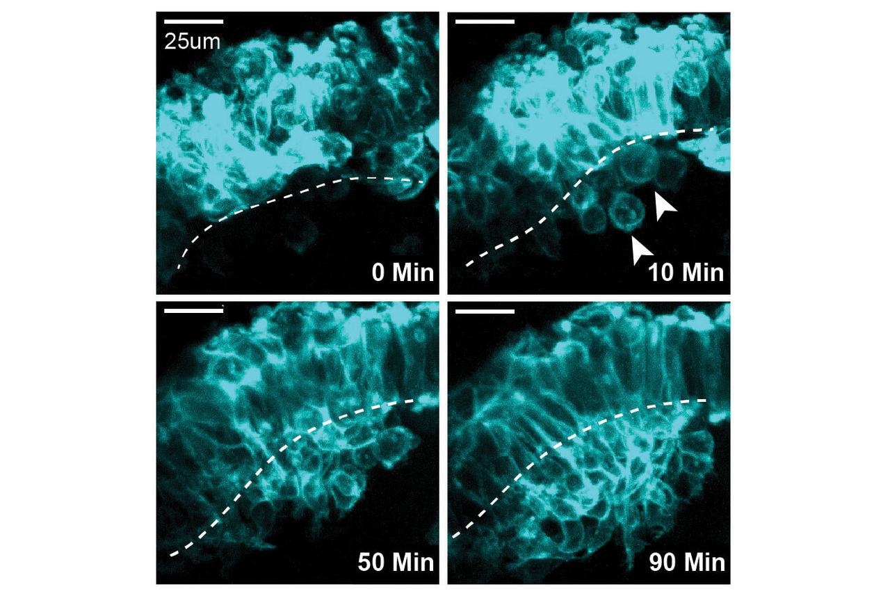
New research from the lab of Stowers Investigator Paul Trainor, Ph.D., has uncovered how mammalian neural crest cells initially detach from their anatomical origin to begin their incredible journey. The study, published in the Proceedings of the National Academy of Sciences on March 10, 2025, and led by Postgraduate Researcher Emma Moore Zajic, Ph.D., a former predoctoral researcher at Stowers Graduate School, used high resolution live imaging to watch neural crest cells mobilize in mice and discovered that a significant percentage of these cells detach differently than expected.
“Neural crest cells have a fascinating developmental journey,” said Moore Zajic. “As the location where they originate is a long way from where they settle, we wanted to understand how these cells first detach and then migrate to generate the bones and cartilage of the head and face.”
Traditionally, neural crest cells were thought to detach from their source solely through a process called epithelial-mesenchymal transition, in which densely packed structural cells called epithelial cells lose their adhesive properties and become migratory. However, Moore observed that in response to enough pressure and tension, neural crest cells can extricate themselves from crowded conditions through cell extrusion, a mechanism often used by cancer cells to detach from tumors.
“Cell extrusion, therefore, is an example of the same cellular processes being used reiteratively during development and disease,” said Trainor.
Moore Zajic used live imaging and staining techniques in transgenic mice to tag and track neural crest cells in real-time and discovered a significant population—between 20-30%—of neural crest cells detached as round cells. Shortly thereafter, these round neural crest cells become mesenchymal, a term used to describe cells that are flexible, able to migrate, and transform into many types of cells.
The research team also identified the molecular mechanism facilitating the extrusion process. A key protein called PIEZO1, which senses tissue pressure and tension, was required for the detachment of this subset of neural crest cells. In addition, mutations in the PIEZO1 gene have been identified in patients with craniofacial anomalies, making this new discovery a significant step toward understanding these conditions.
“Our findings will enable a better understanding of neural crest cells and craniofacial development overall and have the potential for uncovering insights into cancer progression,” said Moore Zajic.
Additional authors include Ruonan Zhao, Ph.D., Mary McKinney, Ph.D., Kexi Yi, Ph.D., and Christopher Wood, Ph.D.
This work was funded by the National Institute for Dental and Craniofacial Research of the National Institutes of Health (NIH) (award: F31DE032256) and with institutional support from the Stowers Institute for Medical Research. The content is solely the responsibility of the authors and does not necessarily represent the official views of the NIH.