In The News


07 January 2026
Investigator Kamena Kostova, named ‘Cell Scientist to Watch’
From the Journal of Cell Science, Investigator Kamena Kostova named a 'Cell Scientist to Watch'
Read Article
Press Release
The findings are a step toward closing the gap on how we could one day deploy regenerative medicine in humans
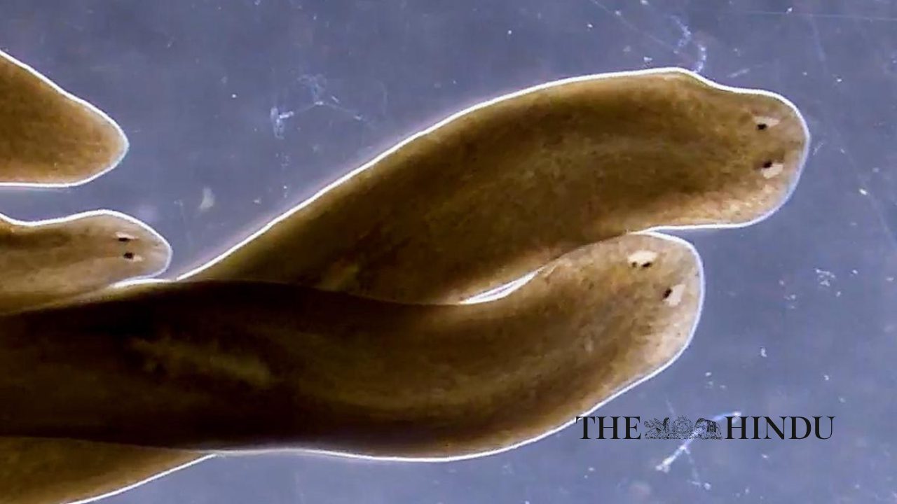
The African killifish
KANSAS CITY, MO—September 26, 2024—Spontaneous injuries like the loss of a limb or damage to the spinal cord are impossible for humans to repair. Yet, some animals have an extraordinary capacity to regenerate after injury, a response that requires a precise sequence of cellular events. Now, new research from the Stowers Institute for Medical Research has unveiled a critical timing factor—specifically how long cells actively respond to injury—involved in regulating regeneration.
A recent study published in iScience on September 20, 2024, sought to understand exactly how an organism knows how much tissue has been lost post-injury. Led by former Predoctoral Researcher Augusto Ortega Granillo, Ph.D., in the lab of Stowers President and Chief Scientific Officer Alejandro Sánchez Alvarado, Ph.D., the team investigated how African killifish properly regrow their tail fin following damage. By analyzing tissue dynamics during regrowth, they found that in addition to known factors, including how many cells are participating and where they are located, the length of time cells spend engaged in the repair process is also key.

The African killifish
“One of the greatest unsolved mysteries of regeneration is how an organism knows what has been lost after injury,” said Sánchez Alvarado. “Essentially, the study points to a new variable in the equation of regeneration. If we can modulate the rate and the length of time that a tissue can launch a regenerative response, this could help us devise therapies that may activate and perhaps prolong the regenerative response of tissues that normally would not do so.”
Shortly after a killifish tail injury, the remaining tissue needs to know how much damage has occurred. Then, this tissue must enlist the right number of repair cells to the site of injury for the right amount of time. Damage sensing, repair cell recruitment, and timing somehow must work together to regrow the tail.
“If an animal that can regenerate extremities, like a tail, loses just a tiny portion, how does it know not to regenerate a whole new tail but just the missing piece?” said Sánchez Alvarado. To address this question, the team probed different locations of injury in the killifish tail fin.

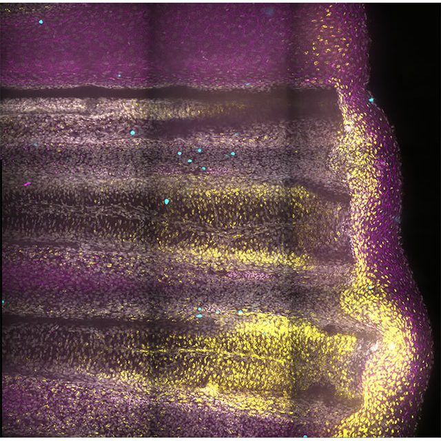
Tail fin regeneration at 6 hours after injury. Fluorescence microscopy image shows skin cells (magenta), actively dividing cells (cyan), and transiently activated skin cell states (yellow).
They found that skin cells both near an injury and in distant, uninjured regions launch a genetic program that primes the whole animal to prepare for a repair response. Then, skin cells at the site of injury sustain this response and temporarily change their state to modify the surrounding material called the extracellular matrix. Ortega Granillo likens this matrix to a sponge that absorbs secreted signals from the injured tissue that then guides repair cells to get to work. If the signals are not received or not interpreted correctly, the regeneration process may not restore the tail’s original shape and size.
“We very clearly defined when and where—at 24 hours post-injury and in the extracellular matrix—the transient cell state is acting in the fin tissue,” said Ortega Granillo. “Knowing when and where to look allowed us to make genetic disruptions and gain a better understanding of the function of these cell states during regeneration.”
To investigate whether these distinct cellular states communicate information to the extracellular matrix—the supportive structure surrounding cells—during the repair process, the researchers employed the CRISPR-Cas9 gene editing technique. They specifically targeted a gene known to modify the extracellular matrix, as they had observed its activation at the onset of the regeneration response. By disrupting the function of this gene, the team aimed to determine its role in relaying information from cells to the matrix during regeneration.
“These modified animals no longer know how much tissue was lost,” said Ortega Granillo. “They still regenerated, but the speed of tissue growth was deficient. This is telling us that by changing the extracellular space, skin cells inform the tissue how much was lost and how fast it should grow.”

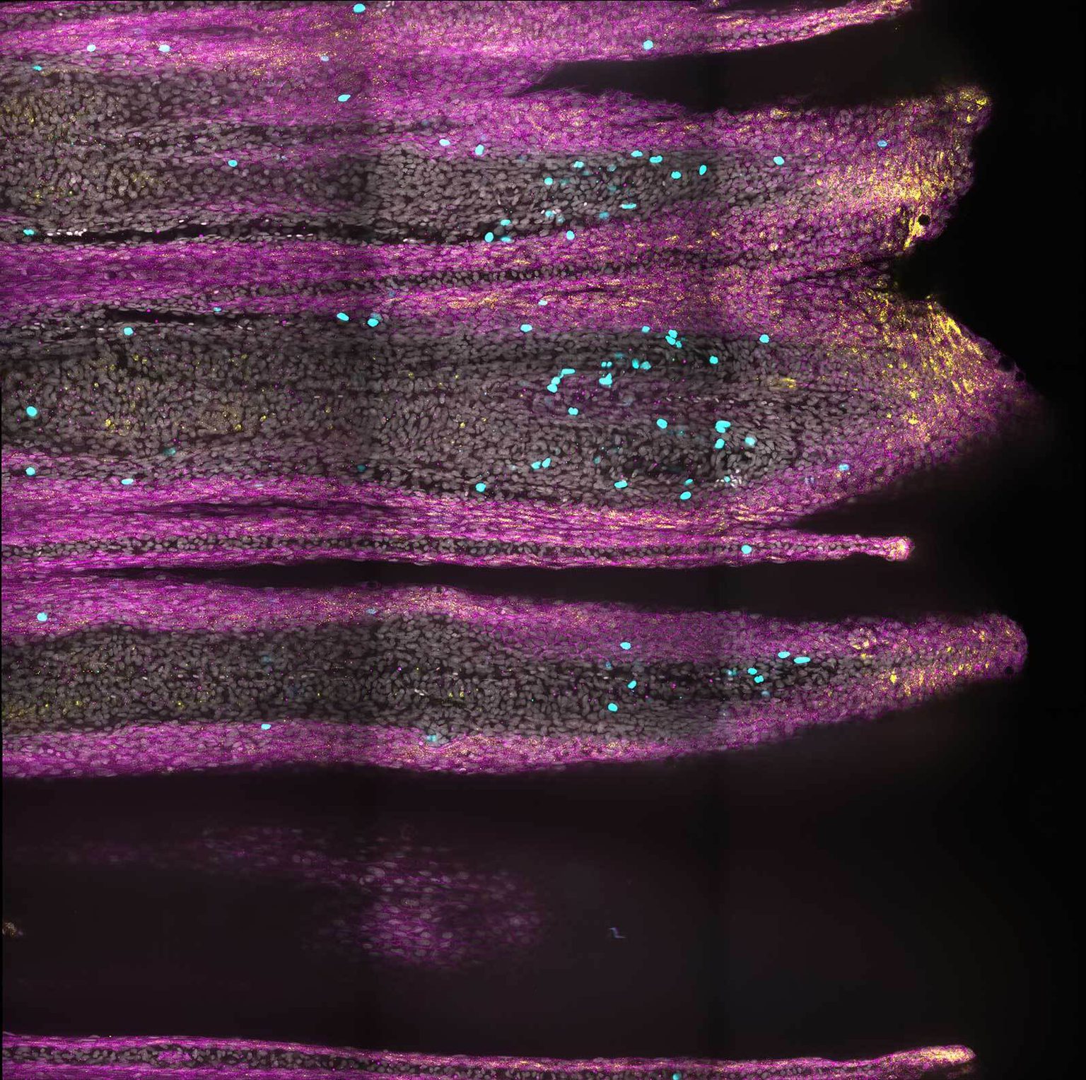
Tail fin regeneration at 7 days post-injury. Fluorescence microscopy image shows skin cells (magenta), actively dividing cells (cyan), and transiently activated skin cell states (yellow), where these cell states are localized now only in the very tip of the fin.
Indeed, the speed and amount of tissue regenerated in these genetically modified killifish increased regardless of whether the tail injury was mild or severe. This finding opens the possibility that cell states that modify the matrix increase regenerative regrowth. If the cell states could be adjusted, it may be a way to stimulate a more robust regeneration response.
From an evolutionary perspective, understanding why certain organisms excel at regeneration while others, such as humans, have limited regenerative abilities is a driving force in the field of regenerative biology. By identifying general principles in organisms with high regenerative capacity, researchers aim to potentially apply these insights to enhance regeneration in humans. This comparative approach not only sheds light on the evolutionary aspects of regeneration but also holds promise for developing novel therapeutic strategies in regenerative medicine.
“Our goal is to understand how to shape and grow tissues,” said Ortega Granillo. “For people who sustain injuries or organ failure, regenerative therapies could restore function that was compromised during illness or following injury.”
Additional authors include Daniel Zamora, Robert Schnittker, Allison Scott, Alessia Spluga, Jonathon Russell, Carolyn Brewster, Eric Ross, Daniel Acheampong, Ning Zhang, Ph.D., Kevin Ferro, Ph.D., Jason Morrison, Boris Rubinstein, Ph.D., Anoja Perera, and Wei Wang, Ph.D.
This work was funded by institutional support from the Stowers Institute for Medical Research and the Howard Hughes Medical Institute.
About the Stowers Institute for Medical Research
Founded in 1994 through the generosity of Jim Stowers, founder of American Century Investments, and his wife, Virginia, the Stowers Institute for Medical Research is a non-profit, biomedical research organization with a focus on foundational research. Its mission is to expand our understanding of the secrets of life and improve life’s quality through innovative approaches to the causes, treatment, and prevention of diseases.
The Institute consists of 21 independent research programs. Of the approximately 500 members, over 370 are scientific staff that include principal investigators, technology center directors, postdoctoral scientists, graduate students, and technical support staff. Learn more about the Institute at www.stowers.org and about its graduate program at www.stowers.org/gradschool.
Media Contact:
Joe Chiodo, Head of Media Relations
724.462.8529
press@stowers.org
In The News
07 January 2026
From the Journal of Cell Science, Investigator Kamena Kostova named a 'Cell Scientist to Watch'
Read Article
#Stowers25: Celebrating 25 Years
06 January 2026
Alejandro Sánchez Alvarado, Ph.D., reflects on a year of discovery, gratitude, and the community that helps support our mission.
Read Article
In The News

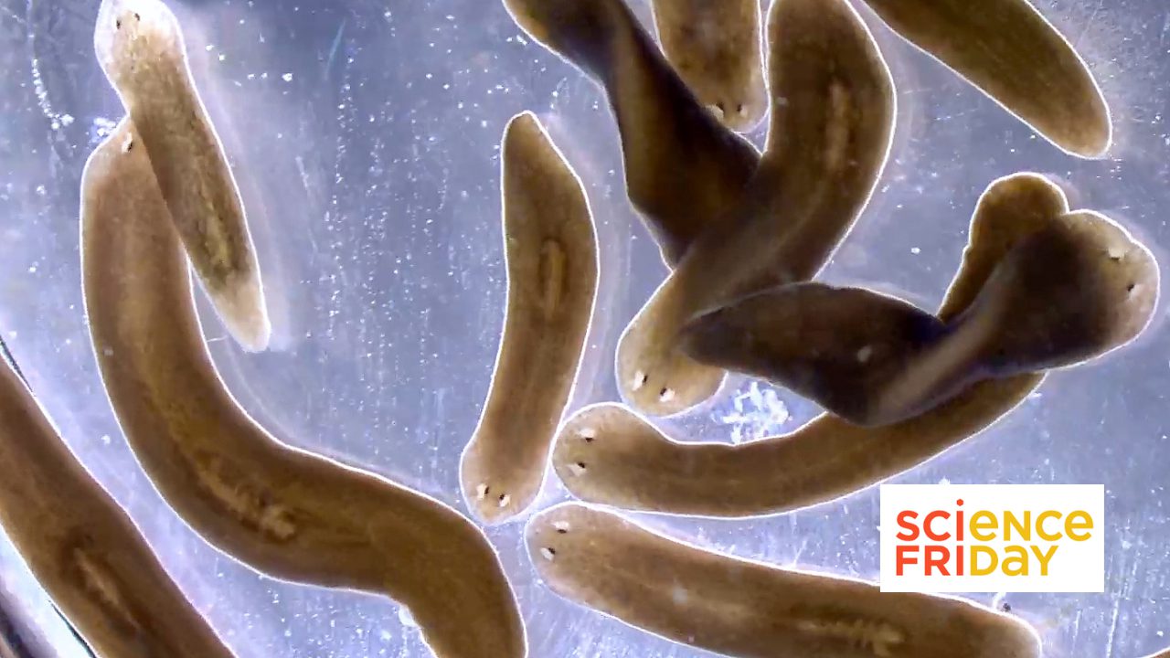
01 January 2026
From Science Friday, President and CSO Alejandro Sánchez Alvarado talks about the science of regeneration and the biology lessons we can carry into the new year.
Read Article