A Stowers Science Visualization Specialist explains the search for seeing cells in a recent journal article.
03 October 2024
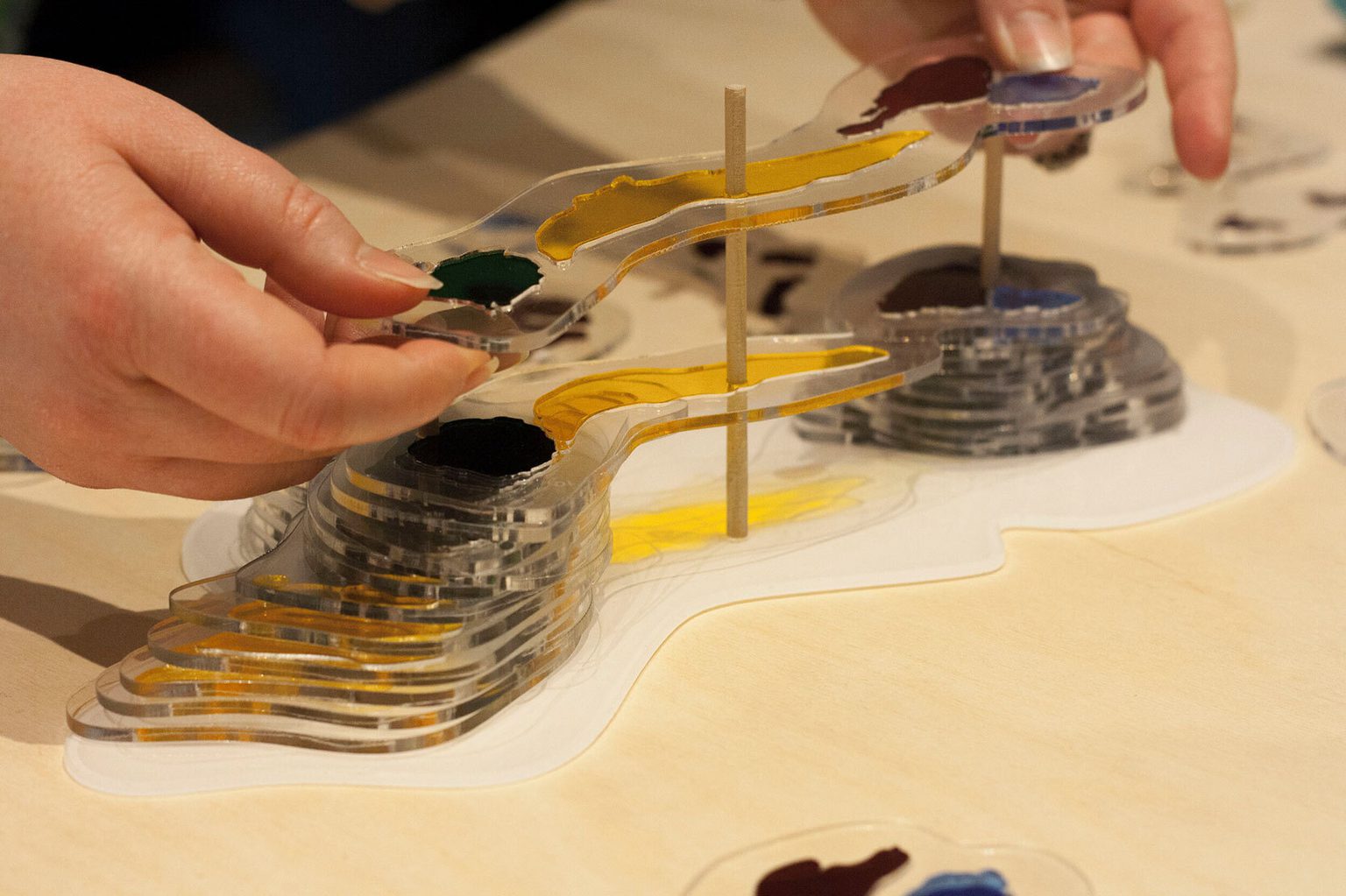
A 3D stacking puzzle for visualizing what cells look like.
Do you remember biology textbook illustrations of cells? Did you understand what cells really look like? That’s the question Mol Mir, a Science Visualization Specialist at the Stowers Institute asks in the opening of a first-person narrative surrounding the search for seeing cells in a recent publication—that also earned the cover image—in the cross-disciplinary journal Patterns.
It began in the summer of 2018 when, as an undergraduate art student struggling to understand what cells looked like and how to convey that picture to others, Mir first visited the Stowers Institute and met Head of Electron Microscopy Stephanie Nowotarski, Ph.D.
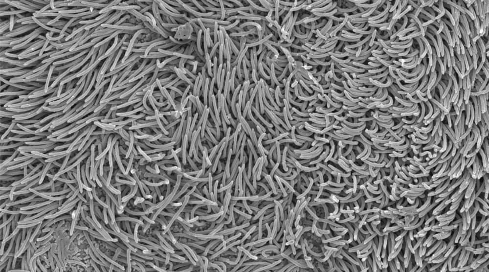
Nowotarski helped Mir see inside of cells, courtesy of science and 3D electron microscopy, while Mir shared artistic insight into 3D printing. Yet Mir was still searching for a tangible way to share this with others.
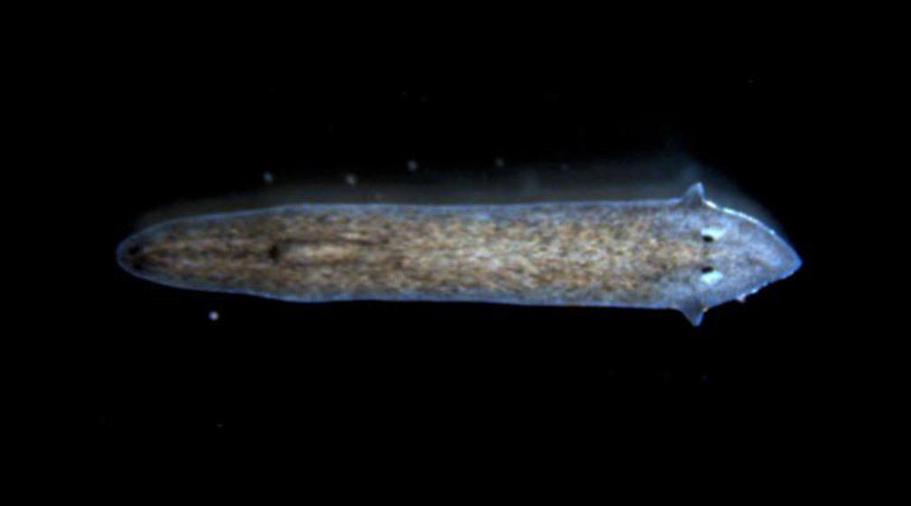
So, Mir turned to flatworms, resolving the 3D structure of their stem cells that allow them to perform astonishing regenerative feats. Like slicing a cabbage and taking a photo of every slice, the deconstruction and reconstruction process allows each layer of a tissue of even a single stem cell to be individually taken apart, visualized under a microscope, and then put back together.
After several attempts at designing methods for interactive visualization, Mir found the right one and manufactured a 3D stacking puzzle, with laser cutting, acrylics, and a keen eye for fun. Mir’s hope is that allowing people to assemble the structure themselves would enable them to understand what cells really look like.
Read more about Mol Mir’s experience bridging art and science.